Spider veins
The secret of the delicate lines
What are spider veins and how do they develop?
Spider veins are dilations of small skin veins that appear as a network of fine lines on the skin, resembling the bristles of a twig broom. They develop when the connective tissue in the vein walls stretches and forms visible, up to 1 mm in size, reddish‑bluish shimmering „bulges".
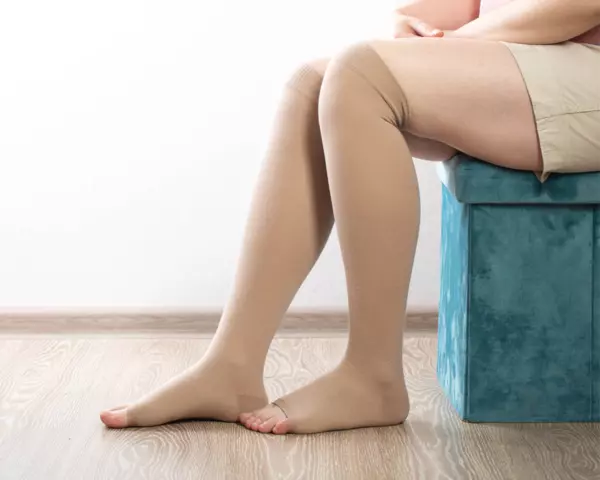
Spider veins are a form of varicose veins (varices, varicosity) and occur particularly frequently on the legs, especially on the thighs, the inner sides of the calves and the ankles.
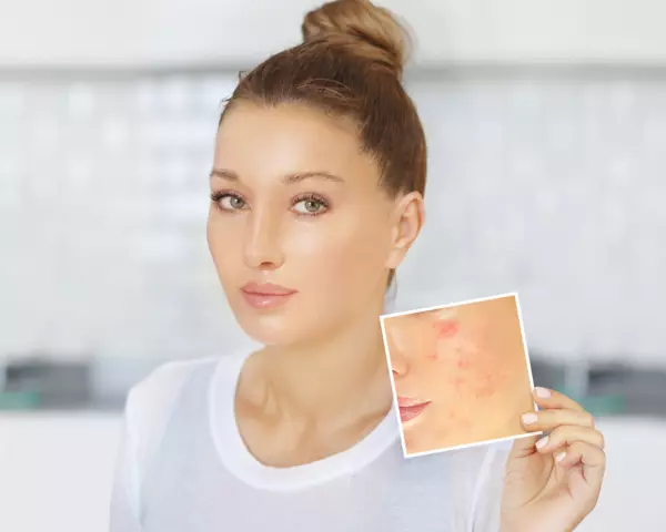
On the face, similar vascular changes are called telangiectasias. They mainly appear on the wings of the nose and above the cheekbones. Unlike spider veins on the legs, they are less often caused by blood backflow and more commonly result from an innate weakness of the connective tissue or age-related vessel dilation. Facial spider veins are generally harmless.
Spider veins and reticular varices share many similarities in appearance and affected areas. Reticular varices are fine, netlike varicose veins that resemble spider veins but are often slightly thicker. Both are normally harmless and do not cause symptoms. Affected people, however, often find the visible veins very bothersome and a detriment to their appearance.
Which factors promote the development of spider veins?
Spider veins can be promoted by various factors. Heredity is likely to play a role. A persistently elevated pressure in the vessels, for example due to high blood pressure, is often responsible for the formation of spider veins.
Prolonged sitting, standing or lying down can hinder blood flow and thus promote the development of spider veins. Overweight and lack of exercise also slow venous blood flow and can cause heavy legs.
Women are more often affected because the female hormone oestrogen relaxes connective tissue and vessel walls. The oral contraceptive pill or a pregnancy can also influence the veins and cause spider veins. Other triggers include nicotine, alcohol and regularly wearing high‑heeled shoes.
Are spider veins dangerous?
Spider veins are mostly harmless and do not cause symptoms. However, they may indicate a weakness of the vein wall that can also affect deeper veins. In particular, with additional symptoms such as heavy legs or swollen ankles, affected individuals should have their venous condition checked by a doctor.
What treatment methods are available for spider veins?
The choice of medical procedure for the treatment of spider veins depends on their size, but costs also play an important role as they vary depending on the method used and the number of sessions required.
In sclerotherapy, also called vein obliteration or sclerotherapy, liquid or foam sclerosing agents are injected directly into the spider veins. These substances irritate and glue the vein walls together, blocking blood flow. Polidocanol is frequently used as the active agent. Treatment costs are relatively low.
In laser therapy, dilated veins are irradiated with laser light of a specific wavelength. The haemoglobin in the blood absorbs the radiation and converts it into heat. This procedure is particularly suitable for very fine spider veins and can be expensive.
Intense Pulsed Light therapy (IPL) resembles laser therapy but works with intense monochromatic light pulses and can also be used for larger spider veins. In radiofrequency therapy, radio waves heat the vein walls, causing the vessels to die off. In electrotherapy or electrocautery, a voltage is applied to the veins via tiny electrodes, causing the vessels to shrink due to heat. This method is considered cost‑effective.
Minimising the risk of spider veins: the most effective tips
- Strengthen your calf muscles with regular exercise such as cycling, running or swimming to promote circulation and prevent pooling in the legs.
- Complement your activities with decongestive and venous gymnastics.
- Avoid prolonged sitting or standing in one place. Make sure to take regular breaks and move to promote blood circulation in the legs.
- Do not cross your legs when sitting to avoid impeding blood circulation.
- Pamper your skin with regular massages, for example under the shower with a brush, to promote circulation and firm the connective tissue.
- Enjoy contrast showers alternating cold and warm water to train vessel elasticity and stimulate blood circulation.
- Maintain a balanced diet with plenty of fresh fruit and vegetables daily, low fat and meat intake, and drink at least two litres of water.
- Avoid smoking in general to reduce the development of spider veins.
- Consider a contraceptive method other than the oral pill to minimise the risk of spider veins.
- Consider using compression stockings to apply gentle external pressure to the veins, which can prevent the formation of spider veins.
- Try home remedies, but always consult your doctor before starting. Make sure you take the right measures to remove spider veins effectively and safely.
- Incorporate plant‑based products such as grape‑leaf extract in the form of creams, capsules or tinctures into your care routine to achieve a possible vein‑protective effect.
- Use horse chestnut extract; its active ingredient aescin improves venous blood circulation. It can be effective in chronic venous insufficiency.
- Rub the affected areas daily with a cloth soaked in apple cider vinegar to benefit from its circulation‑stimulating effect and reduce the appearance of spider veins.
- Support venous blood flow by elevating the legs while sleeping, ideally with special venous pillows. Prevent vessel dilation and reduce the risk of blood pooling to help prevent spider veins.
Spider veins are therefore generally harmless to health, but of course they affect the aesthetic appearance of the skin. They can also be a sign of venous disease, but in any case there are proven means and methods to remove spider veins.
.png.webp)